Are Dental X-Rays Safe and Necessary?
Every year, in the United States, dental X-rays help millions by finding and treating many oral health problems. Despite their common use and big role in keeping mouths healthy, people still often question how safe dental X-rays really are.
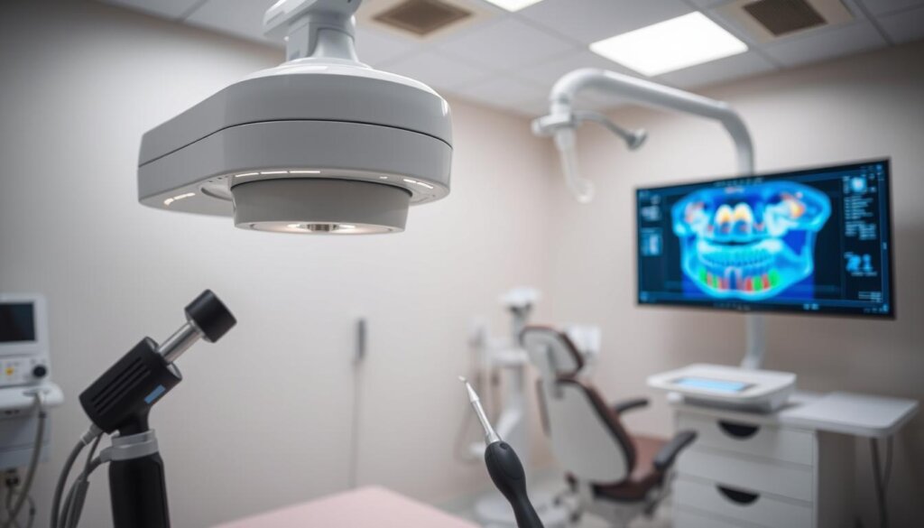
Dental X-rays are key tools for doctors to get a full look inside a patient’s mouth. This look is way beyond what can be seen just by looking. They are crucial in finding and treating dental issues, showcasing the importance of dental X-rays in today’s dentistry. Thanks to dental imaging, doctors can spot hidden issues before they get worse.
Yes, there’s worry about the radiation from X-rays. But, thanks to new dental X-ray safety steps, the risk is now very low. These steps include wearing lead aprons and thyroid collars to keep patients safe. So, dental imaging is not just helpful; it’s made safe with special rules. For details on dental check-ups and cleanings which may include X-rays, check out VisODent NYC.
Key Takeaways
- Many dental X-rays are done safely every year in the U.S., showing they’re widely used and needed.
- The safety precautions for dental X-rays are strict and effective, matching the low radiation exposure.
- The importance of dental X-rays is highlighted by their power to find problems invisible to the eye.
- Professional dental check-ups often include X-rays as part of a complete dental exam and cleaning. More info is available at VisODent NYC.
- Modern improvements in dental imaging keep patient safety first while giving key diagnostic information.
Introduction to Dental X-Rays
Dental X-rays are key in today’s dentistry, showing the health and structure of our teeth and jaws. They help spot various dental problems, such as hidden tooth decay and serious infections in the jawbone. The process uses a little bit of radiation to see differences in how tissues absorb it.
Understanding What Dental X-Rays Are
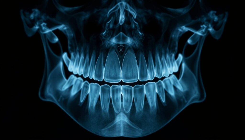
Dental X-rays use a beam of X-ray particles. They go through the mouth to create pictures of teeth, bones, and soft tissues. These images let dentists look beyond the surface, seeing areas not visible in a regular check-up. Thus, they are crucial for full dental care.
Common Types of Dental X-Rays
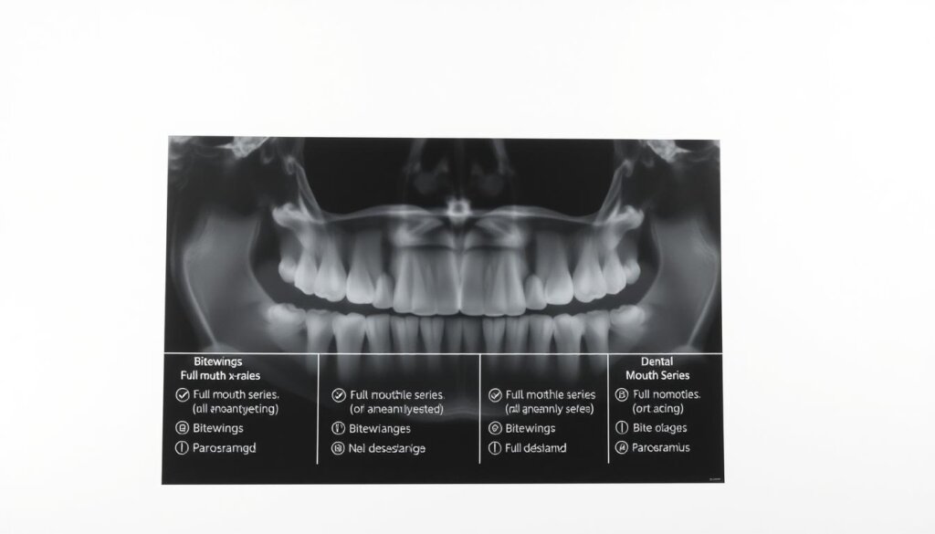
There are two main kinds of dental X-rays: intraoral (inside the mouth) and extraoral (outside the mouth). Intraoral X-rays include bitewing, periapical, and occlusal X-rays. Each has its purpose like checking for cavities or viewing the tooth root. Extraoral X-rays, like panoramic X-rays, show the jaw and skull for assessing teeth alignment and growth.
Purpose of Dental X-Rays in Dentistry
Dental X-rays are vital for preventive health care. They spot problems early, saving patients from pain and complex future treatments. They provide info that helps create an effective treatment plan. This includes finding hidden dental structures, tumors, bone loss, and cavities. Their value in oral health is well acknowledged.
But, it’s important to understand dental X-ray frequency and radiation levels. This knowledge helps lower risks and ensure safety for patients.
The Safety of Dental X-Rays
Dental X-rays are essential in today’s dentistry, helping find and fix dental problems. Yet, some worry about the radiation they give off. It’s important to know how these X-rays are safely used in dental care.
Understanding Radiation Exposure
Dental X-rays do have radiation, but it’s a tiny amount. It’s much less than what we get from nature every year. Think of it like the small amount of radiation you’d get from short plane trips over a few days.
Safety Measures in Dental Imaging
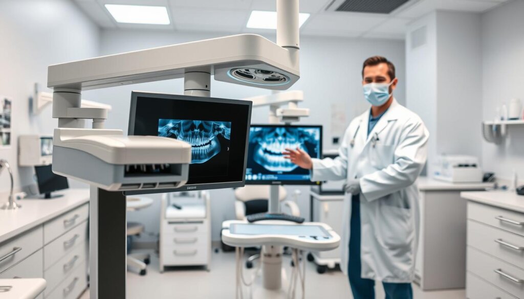
Dentists work hard to keep radiation exposure low. They follow the ALARA principle, which means using as little radiation as possible. Patients also wear protective gear like lead aprons and thyroid collars during X-rays. Plus, digital X-rays now cut down on radiation much more than old film X-rays did.
Comparing Risks of Dental X-Rays to Benefits
It’s important to think about the risks and benefits of dental X-rays. While there is some radiation, the health risk is very low. On the other hand, X-rays can find dental issues early. This can mean easier treatments and better health overall.
Let’s look at how much radiation dental X-rays have compared to other sources:
| Source | Radiation Exposure (in microsieverts) |
|---|---|
| Dental X-rays (per full mouth series) | 150 |
| Airline flights (per hour of flight) | 9 |
| Natural background radiation (yearly average) | 2400 |
In the end, using dental X-rays wisely is key. It helps dentists care for patients better while keeping risks low. If you’re worried about dental X-rays, talk to your dentist. They can explain how it’s safe and why it’s used.
Are Dental X-Rays Necessary?
Dental X-rays are key in assessing oral health, revealing issues not spotted in regular exams. They’re crucial for a complete oral check-up. Dentists use them to see problems hidden from the naked eye. This shows how vital they are in today’s dental care.
When Are Dental X-Rays Recommended?
Dentists decide on X-rays based on each person’s oral health needs. They are crucial during your first visit for a full oral health check. How often you need them after depends on your age, oral health, and disease risk.
Situations That Require Further Imaging
Some oral issues need more detailed images to treat correctly. For instance:
- Spotting decay in tight spaces or under fillings
- Checking gum disease and bone loss
- Preparing for braces by assessing root positions
- Finding cysts or other problems in the jaw
The Role of Dental X-Rays in Preventive Care
Dental X-rays play a big part in stopping dental issues early. They help find decay or gum disease before it’s visible. This early action prevents bigger problems later. It shows how X-rays are a key tool in keeping our mouths healthy.
Dental X-rays are critical for a dentist’s practice. They give a deeper look into oral health, spotting problems early. Understanding their importance can help us see how they keep our teeth healthy. They are essential in both finding and preventing dental issues.
| Issue Detected | Diagnostic Value | Preventive Potential |
|---|---|---|
| Early-stage decay | High | Prevents progression |
| Gum disease | Essential for depth assessment | Limits severity and spread |
| Hidden infections | Critical for location | Facilitates early treatment |
The Types of Dental X-Rays
Dental X-rays are key in helping manage oral health and plan treatments. They let dentists see teeth, roots, how the jaw is placed, and the bones of the face. Dental X-rays come in three main types—Bitewing, Periapical, and Panoramic. Each type is crucial for diagnosing and caring for dental issues.
- Bitewing X-Rays look at how upper and lower teeth line up. They show the tops of back teeth and help find cavities between teeth. They check on fillings and bone density in the jaw, too.
- Periapical X-Rays show the whole tooth, from top to bottom, and the bone around it. They are vital for finding problems with the root and bone, like abscesses or cysts.
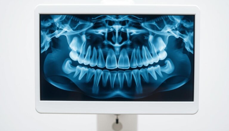
- Panoramic X-Rays give a wide view of the whole mouth, all teeth, jaws, sinuses, and facial bones. They’re great for planning braces, checking for impacted teeth, and finding tumors or breaks.
These X-ray types help dentists diagnose accurately and plan treatments. They also prevent bigger, costlier issues later by catching problems early.
Knowing how and when to use each X-ray type improves patient care. It means treatments are more specific to each person’s needs. This makes sure health and safety are top priorities.
Who Decides on the Need for X-Rays?
The decision on whether you need dental X-rays is made together by you and your dentist. It involves a careful talk that ensures decisions are not only medically right but also agreed by both. This makes sure everyone is on the same page.
Dental provider communication is key to a clear and detailed talk about the need for X-rays. This conversation looks at your health history, and any risks, to decide if X-rays are needed.
Here’s what each person does in deciding if X-rays are necessary:
- The Dentist’s Role in Decision-Making: Dentists check your dental health and past medical records. They then suggest if X-rays are needed to spot problems that can’t be seen during a normal check-up.
- Patient Input and Consent Processes: Your role is also important. You give the go-ahead based on what the dentist tells you. You learn about the good and bad of dental X-rays. This helps you make a well-informed choice on whether to go ahead with it.
Good communication and getting informed patient consent are critical for dental X-ray decision-making. They build trust between you and your dentist. They also make sure dental practices are at their best. Talking with patients about their choices helps dentists make the best use of X-rays. This improves care while keeping you safe and getting good results.
Frequency of Dental X-Rays
Knowing how often to get dental X-rays is key for good oral health. It also helps avoid too much radiation. The rules for this vary a lot. They depend on things like your age, the state of your oral health, and special situations like being pregnant. We’ll look at how often adults, kids, and pregnant women should get dental X-rays. This helps keep their teeth healthy.
Adults: For most adults, getting dental X-rays is part of the usual dental check-ups. But, how often you need them changes based on your mouth’s health, your past with dental decay, or if you’re getting certain dental treatments. It’s really important for dentists to make a custom X-ray plan for each person. This reduces risk and ensures they get the info they need.
Children: Taking care of kids’ teeth is different. Their teeth and jaws change fast, so they might need X-rays more often. This helps watch over their growth and the coming in of new teeth. Dentists must look at each kid’s dental health and growth stage to decide when to do X-rays.
Pregnant Patients: There are extra steps to take care of teeth during pregnancy to keep the baby safe. Even though dental X-rays are safe with the right protections, they’re usually done only if really needed. If you’re pregnant, make sure to tell your dentist. This way, they can make sure both you and your baby are safe.
| Patient Category | Typical Frequency | Special Notes |
|---|---|---|
| Adults | Every 1-2 years | Varies with risk factors |
| Children | Every 6-12 months | Depends on age and dental development |
| Pregnant Patients | Only if essential | Required dental X-rays should use shielding |
This table shows that recommendations on the frequency of dental X-rays change based on the patient. It highlights a way of care that focuses on each person’s health and stage of life.
Risks Associated with Dental X-Rays
Dental X-rays are key in checking oral health, but they have risks. One big worry is dental radiation exposure. Even though it’s small, it could cause dental X-ray side effects.
We’re going to look at the risks of these X-rays. We want to help doctors and patients understand these risks better.
Potential Side Effects of Radiation
Technology has made radiation levels lower. But, getting X-rays often can still be risky. This is especially true for kids and pregnant women. Some dental X-ray side effects are:
- A small increase in cancer risk, like thyroid cancer
- Potential tissue damage from many X-rays over time
- More chance of benign tumors in exposed areas
To lower these risks, it’s important to minimize exposure. Using lead aprons and neck collars helps a lot.
Unintended Diagnoses from X-Rays
Dental X-rays mainly check teeth and gums. But sometimes, they find other health issues. Some accidental discoveries include:
- Early osteoporosis signs
- Hidden jaw tumors or cysts
- Sinus problems
These surprise findings can help in planning better healthcare. They may lead to early treatments that can save lives.
In short, the risks of dental X-rays are low but important to know. Patients and doctors should talk about how often X-rays are needed. They should consider both the risks and the benefits together.
Alternatives to Dental X-Rays
The medical world is always changing, and this includes how we look at teeth. Alternative dental diagnostics are gaining attention because they’re less invasive. Non-radiation dental imaging, like MRI and ultrasound, allows dentists to check your teeth without the risks that come with X-rays.
MRI and ultrasound are leading the way in dental X-ray alternatives. They appeal to people worried about radiation. These options can ease your mind and still give dentists the information they need.
Below, there’s a table that compares these new methods with traditional dental X-rays. It outlines their good points and their downsides. This information can help dentists and patients decide what’s best.
| Technique | Benefits | Limitations |
|---|---|---|
| MRI | No radiation exposure, detailed soft tissue contrast | Higher cost, longer procedure time |
| Ultrasound | Safe for pregnant women, real-time imaging | Lower resolution, limited to certain dental conditions |
| Traditional X-Rays | Low cost, widely available, quick results | Radiation exposure, potential health risks |
Choosing alternative dental diagnostics like MRI and ultrasound lessens radiation risks. They provide different but vital information. While they have their limits and might not replace X-rays fully, their advantages are making them more popular in dentistry today.
Advances in Dental Imaging Technology
The way we look inside our mouths has changed a lot, thanks to digital dental X-rays. This big change is not just a step forward in how we care for our teeth but also opens the door for more improvements. When we think about these changes, it’s key to look at old methods versus new ones and consider what’s next for dental pictures for doctors and their patients.
Digital X-Rays vs. Traditional X-Rays
Dental pros are choosing digital X-rays over old-school film ones for many reasons. Digital X-rays are faster, show clearer images that doctors can adjust to spot issues better, and they don’t expose people to as much radiation—90% less, in fact. Being able to instantly see and share these images makes figuring out and treating dental problems much easier.
The Future of Dental Imaging
The next steps in dental imaging look promising and will change how we diagnose issues. Technologies like cone beam computed tomography (CBCT) now let us make 3D pictures. These detailed images help with more complicated treatments like placing implants. Researchers are also working on ways to use even less radiation without losing the quality of images or making it harder to spot problems.
| Feature | Traditional X-Rays | Digital Dental X-Rays |
|---|---|---|
| Radiation Exposure | Higher | Significantly Lower |
| Image Processing Time | Longer (manual development) | Instant |
| Image Quality | Standard | High; Can be enhanced |
| Diagnostic Applications | Limited to 2D | Extensive (2D and 3D capabilities) |
Cost Considerations and Insurance Coverage
Understanding dental care expenses means knowing how dental X-ray costs work with dental insurance coverage. This part talks about the costs of dental X-rays and insurance policies for these services.
Dental X-ray costs change a lot based on the X-ray type and the dental office’s location. Bitewing X-rays are usually cheaper than panoramic X-rays. We will look into the costs and the insurance plans that could cover them.
| Type of X-Ray | Typical Cost Range | Covered by Insurance? |
|---|---|---|
| Bitewing | $25 – $100 | Yes, mostly as preventive care |
| Periapical | $35 – $150 | Yes, when medically necessary |
| Panoramic | $100 – $250 | Often, particularly for orthodontic treatments |
| Cephalometric projections | $75 – $200 | Specific cases only |
It’s key to review individual insurance benefits when looking at dental procedures. Many plans cover dental X-rays as preventive services, which helps in diagnosis or treatment planning. Yet, insurance plans vary a lot, so knowing the details about your dental insurance coverage can help lower what you pay from your pocket.
Addressing Patient Concerns About Safety
Patients often worry about the safety of dental X-rays. This is due to widespread myths about dental radiation. Talking openly with healthcare providers about these concerns is key. It helps patients understand why dental X-rays are needed and the safety steps taken to protect them.
Understanding and discussing dental radiation safety can significantly reduce patient anxieties and foster a cooperative treatment environment.
Dental professionals need to correct common myths. They should provide clear and factual information to their patients. Doing this can clear up any confusion and increase patient trust in dental practices.
Common Myths About Dental X-Rays
- Dental X-rays cause painful side effects: false, as X-rays are a pain-free imaging technique.
- High radiation exposure levels: in reality, modern dental X-rays involve extremely low levels of radiation.
- Regular X-rays substantially increase cancer risk: extensive studies indicate that the risks are minimal with contemporary imaging technology.
How to Discuss Safety Concerns with Your Dentist
To address dental X-ray myths effectively, consider these steps:
- Prepare your questions: Write down any concerns or questions about dental X-rays beforehand to ensure all your concerns are addressed.
- Ask about advanced technologies: Inquire about the use of digital X-rays, which use up to 90% less radiation than traditional film X-rays.
- Request a review of the results: Ask your dentist to explain the findings and how they impact your treatment plan.
By openly talking about dental X-ray concerns and safety, patients can feel more at ease. They learn about the crucial role these images play in managing dental health.
Understanding the Diagnostic Process
Dental X-rays play a crucial role in accurate dental diagnosis and dental treatment planning. They provide dentists with essential insights. This helps them make treatments precise and minimally invasive.
The heart of this is the dental X-ray diagnostic process. It greatly improves how dentists find and treat mouth health problems not seen in a regular check-up. This type of imaging is key for understanding a patient’s overall dental health and any hidden issues.
How X-Rays Inform Treatment Plans
Dental X-rays are a key tool in creating detailed treatment plans. They show the depth of dental problems like decay, where roots are, and if there are any bone issues. These details are critical for making effective treatment plans. The clear images from X-rays mean every part of a patient’s issue is considered. This leads to care that is both personal and effective.
The Importance of Accurate Diagnosis
Being precise in diagnosing is key for dental treatment to work well. Dental X-rays spot oral health issues early. This avoids the need for bigger treatments later on. This is why dental pros depend so much on these images to make the right care choices.
| Condition Detected | Impact on Treatment Plan |
|---|---|
| Hidden Tooth Decay | Enables targeted decay removal and filling |
| Root Infections | Guides root canal treatment or extraction decisions |
| Bone Loss | Informs the need for bone grafts or other periodontal interventions |
This precise diagnosing not only helps right away but also keeps mouths healthy over time. It shows how vital the dental X-ray diagnostic process is in today’s dentistry.
Case Studies: When X-Rays Made a Difference
Dental X-rays play a huge role in finding problems early. This helps prevent big issues later. By looking at different case studies, we can see the real benefits of getting X-rays on time.
- Early Detection with Dental X-Rays: Case studies show that spotting problems early with dental X-rays leads to better treatments. For example, finding decay between teeth early means less invasive work is needed, avoiding bigger treatments later.
- Consequences of Delayed Dental Imaging: Waiting too long for dental X-rays can make things worse, needing more complex and expensive care. Studies reveal that not doing timely X-rays can let impacted teeth and hidden tumors get to dangerous levels, causing serious health issues.
Looking at case studies helps us understand how important dental X-rays are. They clearly show the bad outcomes of waiting too long for X-rays. They also highlight how catching things early with dental X-rays can prevent future problems.
Conclusion: Balancing Safety and Necessity
In today’s dental world, the balance between balancing dental X-ray safety and necessity is key. We’ve seen how dental X-rays are critical for quality care. They reveal hidden issues like bone loss, cavities, and tumors that we might miss otherwise. Though we worry about radiation, new imaging methods have made it safer.
Dental X-ray final thoughts show how newer technologies have reduced the risks. Today’s dental X-rays use low radiation levels, much less than in the past. Now, patients can understand these risks better and agree to X-rays, knowing they’re vital for preventing dental problems.
Building strong patient-provider communication is vital. This communication helps address any fears patients might have. It’s more than comforting. It strengthens the bond between dentists and patients, helping both. By discussing options together, dentists and patients decide on the best X-ray plans. They focus on the patient’s health and peace of mind.

